(587) 623-6650

Your NE Calgary Choice for Small Parts Ultrasound

Small Parts Ultrasound | Saddletown Radiology | NE Calgary | Radiology Clinic

In the last few decades, there has been a drastic improvement in the technical quality of high-frequency ultrasound for small parts imaging. The addition of colour doppler images has further enhanced the quality of ultrasound imaging. Saddletown radiology is pleased to offer a wide range of diagnostic ultrasound of small parts for all your health needs.

Small Parts Ultrasound | Saddletown Radiology | NE Calgary | Radiology Clinic

What is Small Parts Ultrasound?

Small Parts Ultrasound | Saddletown Radiology | NE Calgary | Radiology Clinic

Small parts ultrasound is used to evaluate possible irregularities in many of the superficial organs of the body such as the thyroid and parathyroid glands, the neck, and the testes. It is a great method of visualizing any swelling, trauma, cysts, nodules, and any other abnormality of soft tissues so that proper diagnosis and a treatment plan can be established. A small parts ultrasound does not use any ionizing radiation; only sound waves to produce images of these areas. No particular preparation for this exam is required.

Thyroid

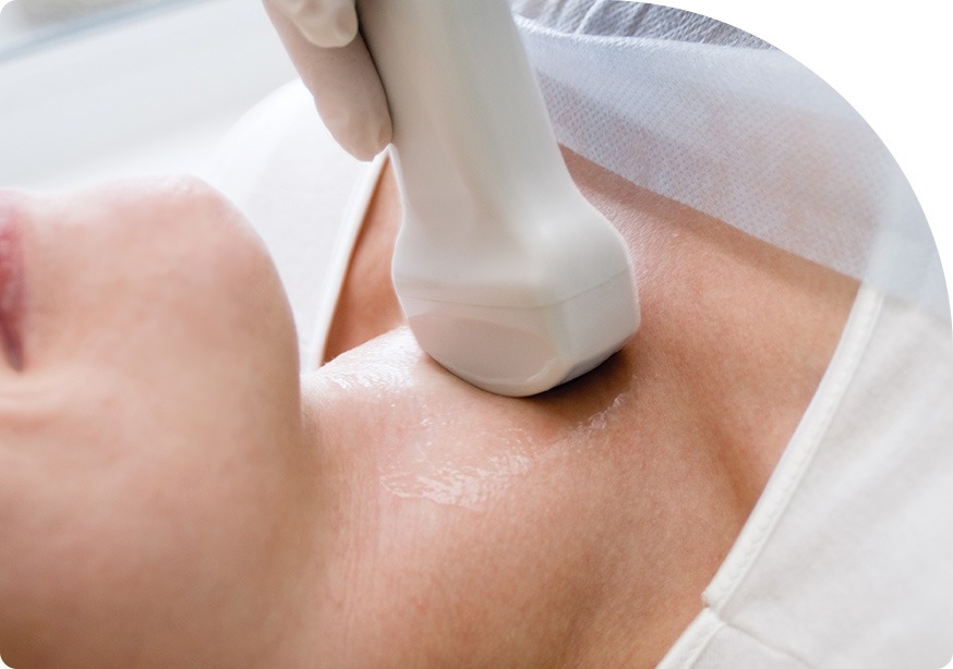
Thyroid Ultrasound | Saddletown Radiology | NE Calgary | Radiology Clinic

Thyroid ultrasound is a popular and useful diagnostic test for evaluating and managing thyroid disorders. Most scans are performed to look at visible or palpable lumps or enlarged glands found during a clinical exam. No preparation is required for this test. During this procedure, you will lie on your back with your neck hyperextended. A clear water-based gel will be used on the neck and thyroid region. A hand-held probe called a transducer will be moved over the regions to produce images that can then be evaluated. 

Thyroid Ultrasound | Saddletown Radiology | NE Calgary | Radiology Clinic
Parathyroid Ultrasound Saddletown Radiology | NE Calgary | Radiology Clinic

Parathyroid

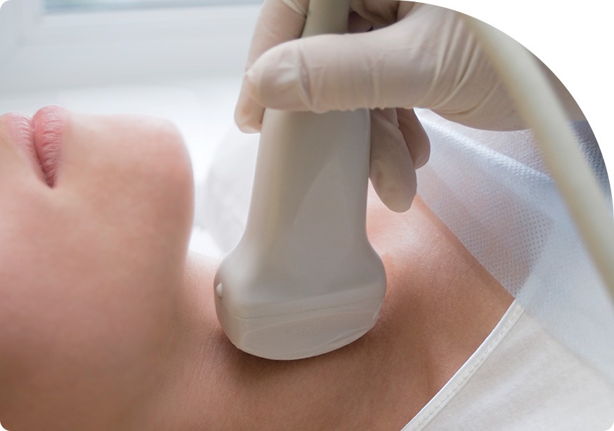
Parathyroid Ultrasound Saddletown Radiology | NE Calgary | Radiology Clinic

A parathyroid ultrasound checks the four parathyroid glands that lie behind or next to the thyroid. The parathyroid makes parathyroid hormone (PTH), which controls the amount of calcium and phosphorus in the blood. A parathyroid ultrasound is done to look for and diagnose enlarged parathyroid glands caused by disease such as parathyroid adenoma. During a parathyroid ultrasound, a small hand-held device called a transducer is passed back and forth over the neck, which takes a picture of the parathyroid glands.

Lymph Nodes

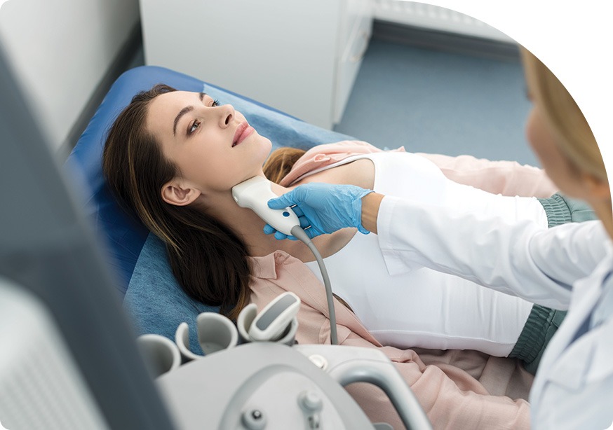
Lymph Nodes Ultrasound | Saddletown Radiology | NE Calgary | Radiology Clinic

A lymph node ultrasound uses high frequency sound waves to create a picture of your lymph nodes. Your lymph nodes are part of the lymphatic system made up of a network of vessels and nodes that carry a clear fluid called lymph around the body. Lymph nodes play a very important part in fighting infection and destroying old or abnormal cells. A lymph node ultrasound is an effective diagnostic tool in the evaluation of inflamed lymph nodes, so that abnormal nodes can be differentiated from healthy ones. If a lymph node ultrasound indicates an irregularity, a biopsy may be performed to determine with certainty the nature of the enlarged nodes. 

Lymph Nodes Ultrasound | Saddletown Radiology | NE Calgary | Radiology Clinic
Salivary Glands Ultrasound Saddletown Radiology | NE Calgary | Radiology Clinic

Salivary Glands

Salivary Glands Ultrasound Saddletown Radiology | NE Calgary | Radiology Clinic

Salivary glands produce saliva in order to moisten the mouth, aid in protecting teeth from decay, and to help digest food. The three major salivary glands include the parotid gland, submandibular gland, and the sublingual glands. An ultrasound of the salivary glands is the first imaging method used for salivary gland swelling. Viral infection is the most common cause of salivary gland swelling, although there are other causes. An ultrasound uses sound waves and helps to see salivary gland cancer and other abnormalities.

Parotid Gland

Parotid Gland Ultrasound | Saddletown Radiology | NE Calgary | Radiology Clinic

The parotid glands are the two largest salivary glands located just in front of the ears on either side of the face. These glands produce saliva to help with chewing and digesting food. Most salivary gland tumors start in the parotid gland. Viral infections like the mumps, the flu, and other kinds can cause swelling of glands. A parotid gland ultrasound uses sound waves to create images of the parotid glands and helps to see the glands and determine the cause of any swelling. While the lobe of the parotid is deep, and can be difficult to see using ultrasound, ultrasound is still the initial diagnostic procedure to evaluate changes of the parotid gland; it has a higher accuracy in delineating benign tumours from malignant salivary gland tumours.

Parotid Gland Ultrasound | Saddletown Radiology | NE Calgary | Radiology Clinic
Parathyroid Ultrasound | Saddletown Radiology | NE Calgary | Radiology Clinic

Submandibular Gland

Parathyroid Ultrasound | Saddletown Radiology | NE Calgary | Radiology Clinic

The submandibular gland is the second largest of the three main salivary glands (which includes the parotid and sublingual gland), and lies deep in the body of the mandible, protruding slightly and superficially. The function of the submandibular glands is to lubricate and moisturize the mouth and the throat for digestion. Sometimes the submandibular glands can become swollen or enlarged, usually due to tiny stones blocking the ducts that channel saliva into the mouth. Sometimes however, swelling can be caused by the presence of cancer. When there is pain, this is sometimes confused with an inflammatory disorder. Less common signs of submandibular gland cancers include tumor fixation, skin invasion, lower face paralysis, and enlarged neck nodes.

Neck

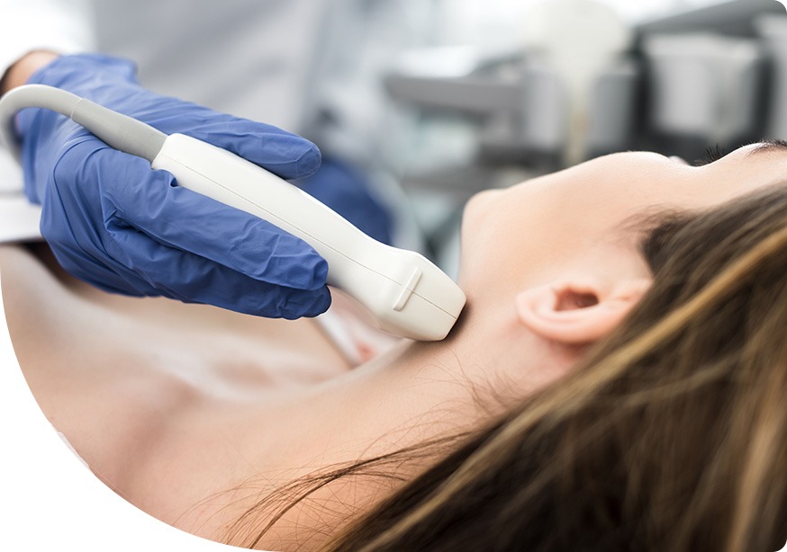
Neck Ultrasound Saddletown Radiology | NE Calgary | Radiology Clinic

A neck ultrasound can be used to evaluate any potential problems of the thyroid, lymph nodes, and carotid arteries, located on each side of the neck. The arteries in the neck deliver blood from your heart to your brain. A neck ultrasound uses high frequency sound waves to create picture images from inside the body of the face and neck, and it looks for any growths, nodules, or tumors. A neck ultrasound is safe and generally painless; there is minimal pressure as the transducer is pressed against the area to be examined.

Neck Ultrasound Saddletown Radiology | NE Calgary | Radiology Clinic
Testicular Scrotum Ultrasound | Saddletown Radiology | NE Calgary | Radiology Clinic

Testicular/Scrotum

Testicular Scrotum Ultrasound | Saddletown Radiology | NE Calgary | Radiology Clinic

A testicular/scrotum ultrasound is a diagnostic test used to obtain images of the testicles and surrounding tissues in the scrotum. It is commonly used when a male patient feels pain or an abnormal lump. It can diagnose suspected abnormalities of the scrotum and is the main technique used to evaluate problems of the testicles and surrounding tissue. No preparation is required. A handheld device, called a transducer, that is covered in gel, will be moved back and forth across the testicles /scrotum to obtain the required pictures to be evaluated by the radiologist. A testicular/scrotum ultrasound a painless and non-invasive test.

Epididymis

Epididymis Ultrasound | Saddletown Radiology | NE Calgary | Radiology Clinic

The epididymis is located adjacent to the testis, within the scrotal sac. Its primary function is to collect, mature, and transport sperm via the ductus deferens. Acute epididymitis is the most common condition that causes severe scrotal pain in adults, although there are other reasons. Males of any age can get epididymitis. In order to determine the proper treatment for an epididymis condition, an ultrasound is useful. This is because the imaging may be helpful to see if the blood flow to the testicles is lower than normal, indicating twisting, or higher than normal, which would help confirm the diagnosis of epididymitis.

Epididymis Ultrasound | Saddletown Radiology | NE Calgary | Radiology Clinic

We look forward to hearing from you soon! Call Saddletown Radiology at (587) 623-6650 to book your appointment today!

Read Our Reviews On

Google Logo

4.9

review-star-icon

Contact Saddletown Radiology

Name(Required)

Our Hours

  • Monday: 8:00 am to 5:30 pm
  • Tuesday: 8:00 am to 5:30 pm
  • Wednesday: 8:00 am to 5:30 pm
  • Thursday: 8:00 am to 5:30 pm
  • Friday: 8:00 am to 5:30 pm
  • Saturday: Closed
  • Sundays: Closed
  • Statutory Holidays: Closed

Providing Radiology Services to the NE Calgary communities of Saddle Ridge, Martindale, Taradale, Castleridge, Falconridge, Coral Springs, Westwinds, Cityscape, Stoney & Surrounding Calgary Areas

We Accept All Requisitions From Any Clinic | Saddletown Radiology | NE Calgary | Radiology Clinic