(587) 623-6650

Your NE Calgary Choice for all your General Ultrasound Needs

General Ultrasound | Saddletown Radiology | NE Calgary | Radiology Clinic

Saddletown Radiology is pleased to provide comprehensive general ultrasound services to meet all your health needs. General ultrasound imaging uses sound waves to produce pictures of the inside of the body, displaying the images in thin, flat sections of the body. These images help examine many of the body’s internal organs and can help to diagnose the cause of things like swelling, pain, and infection in the body’s internal organs, including the gallbladder, liver, kidneys, uterus, pancreas, ovaries, prostate, testicles, breasts, thyroid, and some musculoskeletal tissues. General ultrasound can also be used to examine an unborn child (fetus) in pregnant women, as well as to evaluate the blood flow in the veins and arteries in the neck, legs, abdomen, and heart. General ultrasound is safe, noninvasive, and does not use radiation.

General Ultrasound | Saddletown Radiology | NE Calgary | Radiology Clinic
Abdominal Ultrasound | Saddletown Radiology | NE Calgary | Radiology Clinic

Abdominal Ultrasound

Abdominal Ultrasound | Saddletown Radiology | NE Calgary | Radiology Clinic

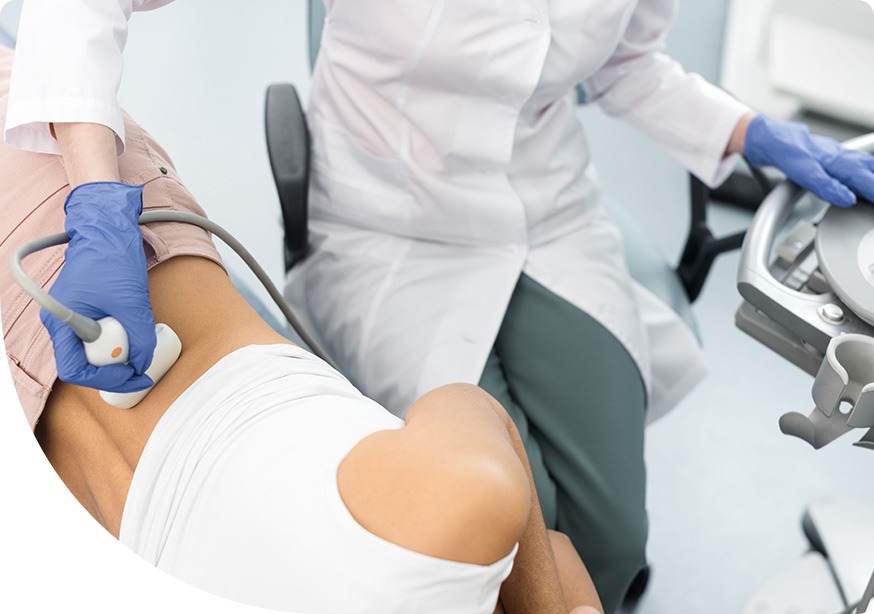
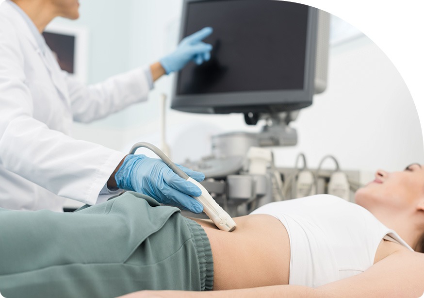
An abdominal ultrasound uses invisible sound waves to produce images of internal tissue and organs. It is often performed in order to assess and evaluate the abdominal area for causes of abdominal pain, or to assess the anatomy of the abdominal organs. Abdominal ultrasounds look at things including the liver, pancreas, gallbladder, spleen, bile ducts, and abdominal aorta. During the Abdominal ultrasound, the machine sends sound waves into the body tissues, and then black and white images are recorded on a computer screen. These images show the internal structure of the abdominal organs for evaluation by a Radiologist.

Renal Ultrasound

Renal Ultrasound | Saddletown Radiology | NE Calgary | Radiology Clinic

A renal ultrasound looks at one or both kidneys using a diagnostic ultrasound. The kidneys filter waste products out of your blood, which then leave the body as urine. A detailed picture of the kidneys is important in the diagnosis and management of kidney-related diseases. This noninvasive diagnostic exam produces images of the kidney that are used to assess the shape, size, and location of the kidneys and related structures, or to assess the blood flow to the kidneys. Renal ultrasounds can detect things like tumors, abscesses, cysts, obstructions, fluid collection and infection within the kidneys or around the kidneys.

Renal Ultrasound | Saddletown Radiology | NE Calgary | Radiology Clinic
Bladded Ultrasound | Saddletown Radiology | NE Calgary | Radiology Clinic

Bladder Ultrasound

Bladded Ultrasound | Saddletown Radiology | NE Calgary | Radiology Clinic

A bladder ultrasound is a noninvasive and generally painless test that uses sound waves to create a picture of what is inside the bladder, providing information about the wall of the bladder, diverticula (pouches) of the bladder, bladder stones, and large tumours in the bladder. Bladder ultrasounds are ordered when there is concern about bladder problems, such as difficulty urinating or daytime wetting. A bladder ultrasound is able to show how much urine the bladder holds when it is full, and whether someone completely empties the bladder when urinating. A full bladder is very important for the exam. Patients are asked to consume one litre of fluid about an hour before the exam.

Vascular Ultrasound

Vascular Ultrasound | Saddletown Radiology | NE Calgary | Radiology Clinic

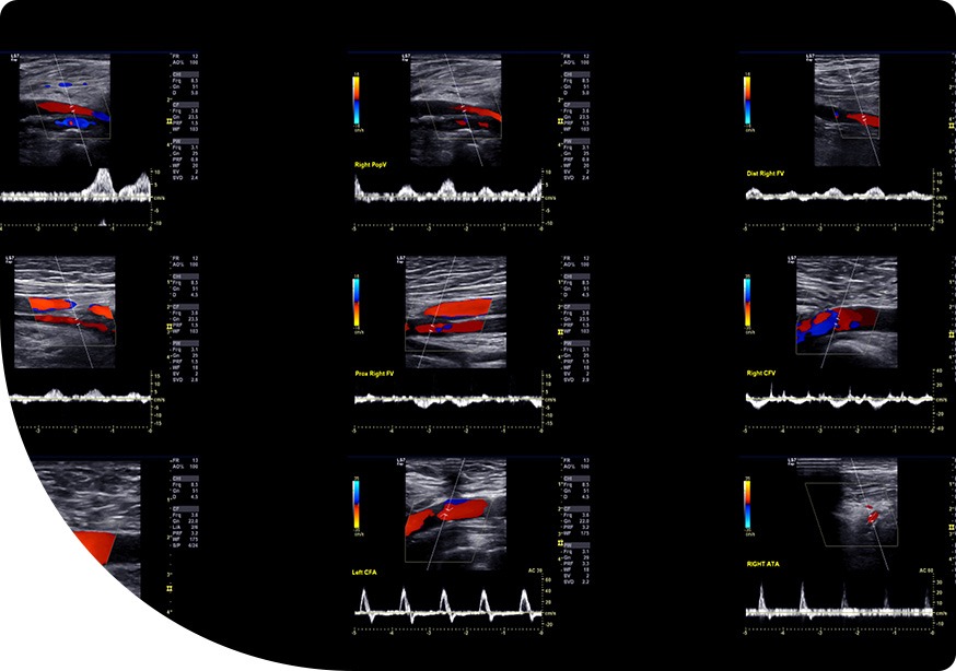
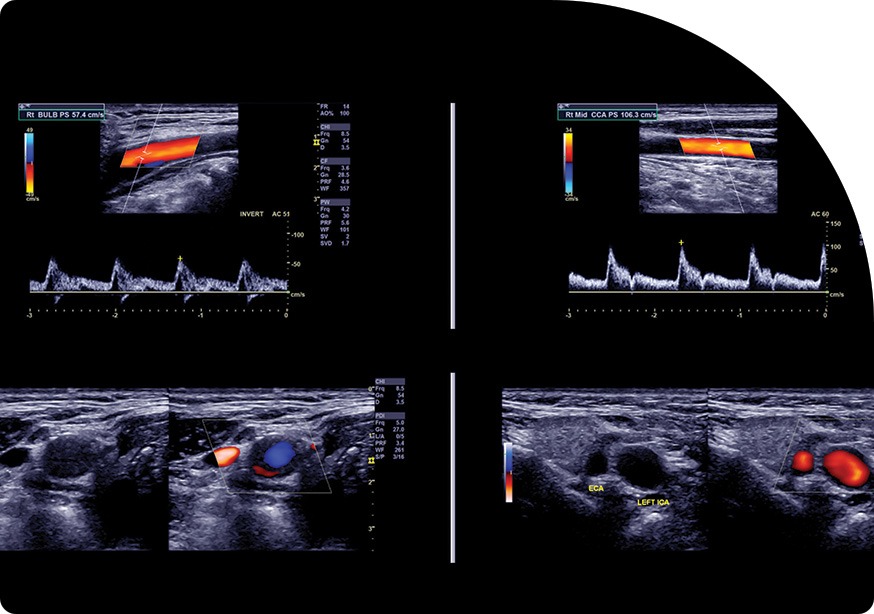
Vascular ultrasound is an ultrasound method used to examine the circulation in the body’s blood vessels. It can also evaluate veins or arteries in nearly any part of the body. It may be recommended in order to locate and identify blockages and abnormalities, detect blood clots in major veins of the legs or arms, determine whether a patient is a good candidate for a vascular procedure, determine if there is an enlarged artery (aneurysm), or evaluate varicose veins, among other things. Evaluating circulation helps determine if there is a risk of stroke or vascular disease. A vascular ultrasound is non-invasive, meaning it does not require the use of dyes, needles, radiation, or anesthesia. During a vascular ultrasound, sound waves are transmitted through the tissues of the area being examined, which reflect off blood cells moving within the blood vessels. The sound waves then return to the ultrasound machine, where they are recorded and displayed on a computer screen as an image of the blood vessel. The findings allow for the calculation of the speed at which the blood cells are moving.

Vascular Ultrasound | Saddletown Radiology | NE Calgary | Radiology Clinic
Arterial Ultrasound | Saddletown Radiology | NE Calgary | Radiology Clinic

Arterial Ultrasound

Arterial Ultrasound | Saddletown Radiology | NE Calgary | Radiology Clinic

An arterial ultrasound uses sound waves to create a colour map of the arteries in order to identify narrowing of your vessels that may be causing pain. An arterial ultrasound may help to diagnose various conditions such as blood clots, damaged or dysfunctional valves in the veins in your legs, or defects of your heart. The process takes about 20-30 minutes for each arm or leg imaged. A water-soluble gel is placed on a handheld device called a transducer. The transducer directs high-frequency sound waves to the artery or veins being tested. Blood pressure cuffs may be put around different parts of the body, including the calf, thigh, ankle, and different points along the arm. Once the colour map of the arteries is created, a radiologist is able to examine it for any signs of narrowing vessels.

Hernia Ultrasound

Hernia Ultrasound | Saddletown Radiology | NE Calgary | Radiology Clinic

A hernia occurs when an area of tissue or organ pushes through the groin or abdominal muscle. If left untreated, hernias can lead to severe complications, so it is important to schedule an appointment with an experienced specialist who can quickly diagnose and treat your hernia. Sometimes a hernia can be diagnosed with a physical exam only. Other times, there may be symptoms of a hernia (like pain), but no obvious protrusion or palpable mass. In such cases, a hernia ultrasound may be ordered. Using a handheld transducer, a hernia ultrasound is a noninvasive and simple test that uses sound waves to create real-time images of the precise location of your pain and/or the locations most commonly associated with hernia, in order to definitively diagnose a hernia and seek the appropriate treatment.

Hernia Ultrasound | Saddletown Radiology | NE Calgary | Radiology Clinic
Obstetrics/Pregnancy Ultrasound | Saddletown Radiology | NE Calgary | Radiology Clinic

Obstetrics (Pregnancy)

Obstetrics/Pregnancy Ultrasound | Saddletown Radiology | NE Calgary | Radiology Clinic

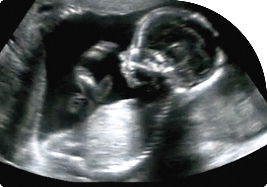
Obstetrics is a branch of medicine focusing on all aspects of pregnancy, from prenatal care and support during pregnancy, to childbirth and postpartum care. Obstetricians have specialized skills to deal with complex or high-risk pregnancies and birth, and they are qualified to perform interventions and caesareans if needed. They take care of you throughout your pregnancy and can provide follow-up care such as annual Pap tests for years to come. Saddletown Radiology is equipped to provide the unique and diagnostic tests required of mothers-to-be and their unborn children, including obstetric ultrasound, which uses high-frequency sound waves to produce images of the developing embryo or fetus. An obstetric ultrasound is also able to monitor the health of the mother’s ovaries, uterus, and the blood flow through the umbilical cord to the placenta. Using the findings from obstetrics ultrasounds, doctors are able to track the progress of your pregnancy, gestational age, and help to predict delivery dates.

Vascular Doppler Ultrasound (DVT & Arterial Ultrasound)

Vascular Ultrasound | Saddletown Radiology | NE Calgary | Radiology Clinic

A vascular doppler ultrasound is a noninvasive test that evaluates movement of materials in the body. While a regular ultrasound uses sound waves to produce images, it cannot show blood flow. A vascular doppler ultrasound uses sound waves to evaluate the body’s circulatory system, and helps identify blockages in the arteries and veins, as well as detect blood clots. Seeing and evaluating blood flow is helpful to detect blood clots in the vein, confirm leaky valves in the veins that can cause swelling or edema, and confirm chronic venous insufficiency. You may need a vascular doppler ultrasound if you have any symptoms of reduced blood flow or heart disease, including weakness or numbness in your legs, a cold feeling in your lower foot or leg, cramping that is painful in your leg muscles or hips when climbing or walking stairs, or a change in colour and/or shiny skin on your leg. You may also need a vascular doppler ultrasound if you have had an injury to your blood vessels, have suffered a stroke, are being treated for a blood disorder, or are pregnant and it is thought that your or your unborn baby might have a blood flow problem.

Vascular Ultrasound | Saddletown Radiology | NE Calgary | Radiology Clinic
Abdominal Wall Ultrasound | Saddletown Radiology | NE Calgary | Radiology Clinic

Abdominal Wall

Abdominal Wall Ultrasound | Saddletown Radiology | NE Calgary | Radiology Clinic

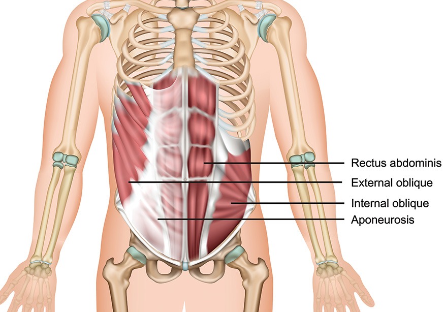
An abdominal wall ultrasound is a diagnostic procedure that uses sound waves to create a picture of the abdominal wall. Using a handheld probe that glides over a special gel applied to the testing area, the sound waves produced are translated into pictures of the soft tissues inside of the body. These pictures help physicians evaluate the structures of the abdominal wall and diagnose medical conditions. An abdominal wall ultrasound is a safe and noninvasive test that can check for many conditions.

Pelvic Ultrasound

Pelvic Ultrasound | Saddletown Radiology | NE Calgary | Radiology Clinic

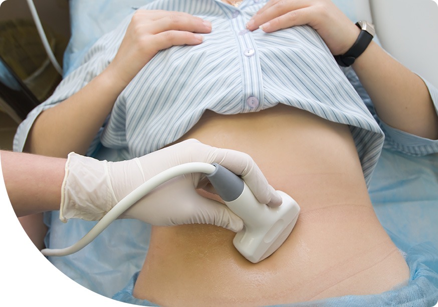
A pelvic ultrasound is a safe and noninvasive diagnostic exam that uses sound waves to produce images that are used to evaluate organs and structures within the pelvis. Using a handheld device called a transducer, that is placed on the abdomen using a conductive gel, a pelvic ultrasound in women would include investigation of the cervix, fallopian tubes, ovaries, uterus, vagina, and bladder. In men, it would include the bladder, the prostate, and the seminal vesicles. A pelvic ultrasound can help diagnose and assist in the treatment of any abnormalities in the structure of the uterus, like cysts or tumours, and would include endometrial conditions. In terms of preparation for a pelvic ultrasound, your bladder will need to be full, which makes the organs show up more clearly in the pictures for easier reading.

Pelvic Ultrasound | Saddletown Radiology | NE Calgary | Radiology Clinic
Endovaginal Ultrasound | Saddletown Radiology | NE Calgary | Radiology Clinic

Pelvic Ultrasound with Endovaginal scan

Endovaginal Ultrasound | Saddletown Radiology | NE Calgary | Radiology Clinic

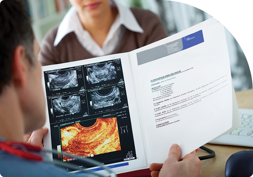
A pelvic ultrasound is a safe noninvasive exam that produces pictures that are used to assess organs and structures within the female pelvis, using a handheld device called a transducer, which is placed on the abdomen using conductive gel. A transabdominal exam is able to offer a wide view of the pelvic organs. The Endovaginal scan includes a thin transducer cover with a lubricated sterile protection that is placed in the vagina, much like a tampon, to examine the reproductive organs. An Endovaginal ultrasound is able to provide far greater detail in a smaller area. Sometimes, a transabdominal ultrasound will be initially performed and then followed by an Endovaginal exam if it is necessary to have more detailed images. With pelvic ultrasound, your bladder needs to be full. However, with the Endovaginal scan, your bladder needs to be empty, so you will use the bathroom after your pelvic ultrasound and before the Endovaginal scan.

We look forward to hearing from you soon! Call Saddletown Radiology at (587) 623-6650 to book your appointment today!

Read Our Reviews On

Google Logo

4.9

review-star-icon

Contact Saddletown Radiology

Name(Required)

Our Hours

  • Monday: 8:00 am to 5:30 pm
  • Tuesday: 8:00 am to 5:30 pm
  • Wednesday: 8:00 am to 5:30 pm
  • Thursday: 8:00 am to 5:30 pm
  • Friday: 8:00 am to 5:30 pm
  • Saturday: Closed
  • Sundays: Closed
  • Statutory Holidays: Closed

Providing Radiology Services to the NE Calgary communities of Saddle Ridge, Martindale, Taradale, Castleridge, Falconridge, Coral Springs, Westwinds, Cityscape, Stoney & Surrounding Calgary Areas

We Accept All Requisitions From Any Clinic | Saddletown Radiology | NE Calgary | Radiology Clinic